

Pelvic Ultrasound in Kondapur, Hyderabad
A pelvic ultrasound is a simple, painless scan that gives your doctor a clear picture of your uterus, ovaries, and surrounding pelvic organs. There is no radiation involved, and in most cases, you get your results the same day.
At Matrika Fertility and Laparoscopic Centre, a trusted IVF center in Kondapur, Hyderabad, Dr. Pooja Papishetty personally reviews every pelvic ultrasound. This isn’t a scan you get done at one place and interpreted at another. The person who understands your history is the same person reading your scan.
“A pelvic ultrasound often tells us more in ten minutes than weeks of guesswork. It’s usually the first real step toward answers.” – Dr. Pooja Papishetty, Gynaecologist and Fertility Specialist, Kondapur, Hyderabad
What is a Pelvic Ultrasound?
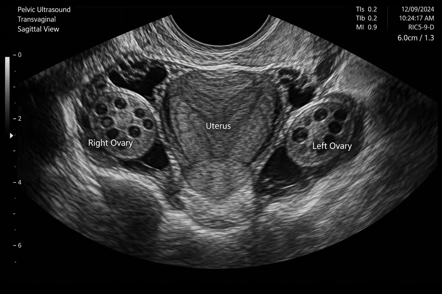
A pelvic ultrasound, commonly referred to as a gynaecological USG, uses sound waves to create real-time images of the organs inside your pelvis. This includes the uterus, ovaries, fallopian tubes, cervix, and bladder.
It’s one of the most commonly used diagnostic tools in gynaecology and fertility care because it’s quick, non-invasive, and doesn’t involve any radiation. Unlike an X-ray or CT scan, it’s considered completely safe, even during pregnancy.
As a fertility scan, it plays a central role in tracking ovulation, checking uterine health, and monitoring follicle growth during treatment cycles. Doctors also use it to check the size and shape of reproductive organs, look for cysts or fibroids, or confirm an early pregnancy.
Types of Pelvic Ultrasound
| Type | How It’s Done | Best For |
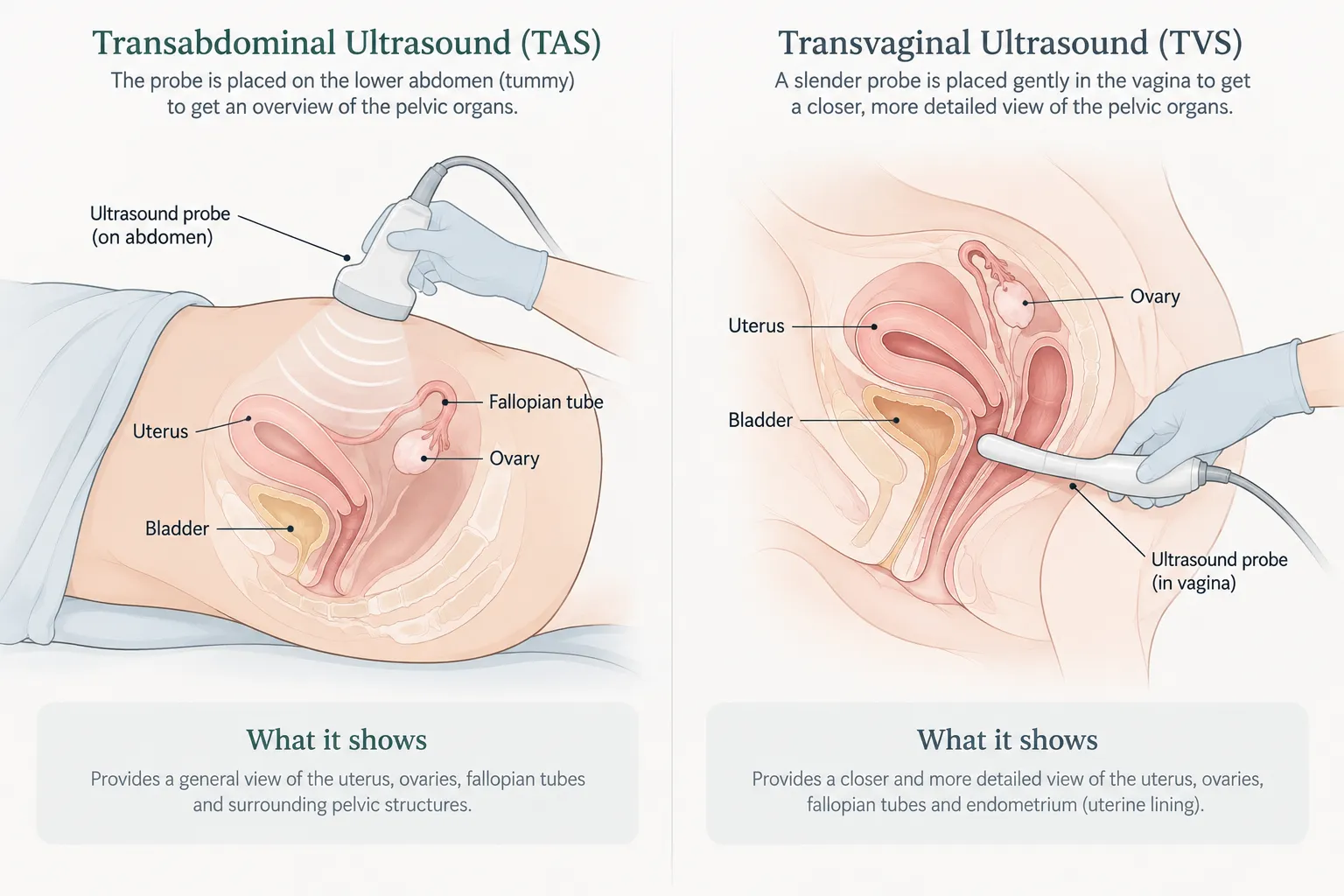
| Transabdominal Ultrasound | Probe moved over the lower abdomen with gel | General pelvic organ view, full bladder needed |
| Transvaginal Ultrasound | Slim probe inserted into the vagina | Detailed view of uterus, ovaries, and follicles
|
| 3D Pelvic Ultrasound | 3D probe, usually done transvaginally | Uterine shape, structural defects, and fertility evaluation |
What Is a 3D Pelvic Ultrasound and When Is It Used?
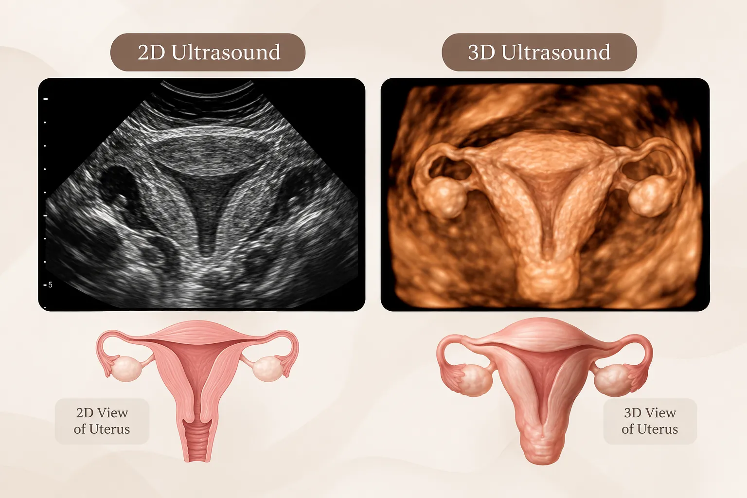
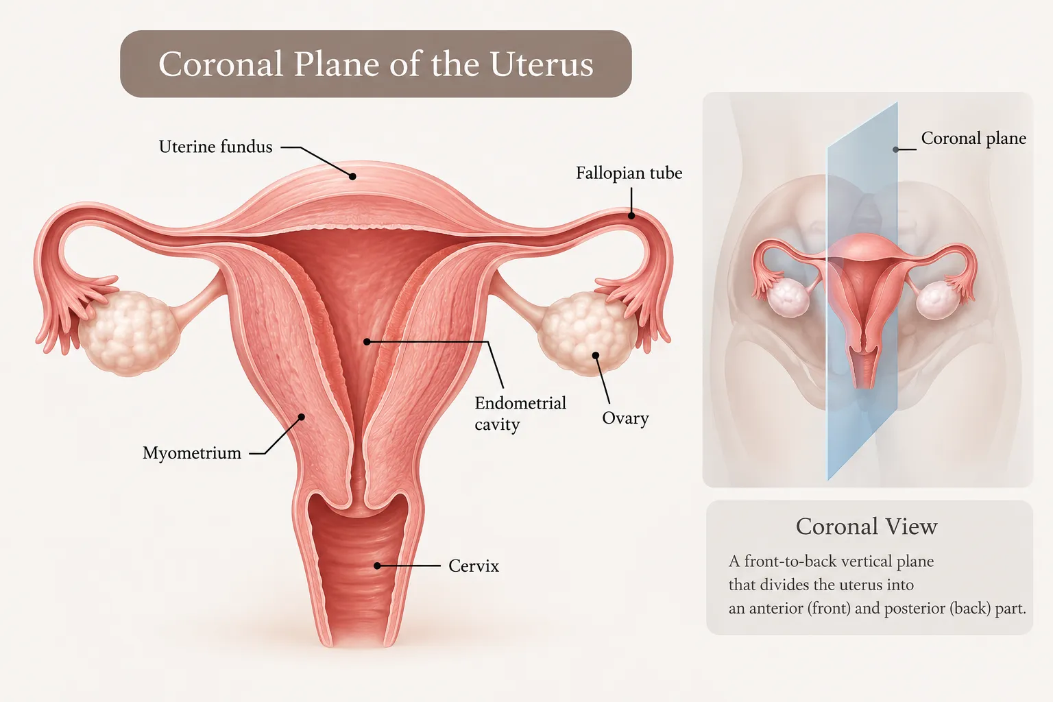
A regular 2D scan shows the pelvic organs as flat, cross-sectional images. A 3D ultrasound combines many of these images into one three-dimensional view. This helps the doctor see the uterus from more angles, including the coronal view, which a 2D scan cannot show clearly.
A 3D scan is not needed for every patient. Doctors usually suggest it to:
● Check the shape of the uterus and spot a septum or other structural variation
● Locate fibroids or polyps more precisely
● Assess the uterine lining
● Count antral follicles as part of a fertility evaluation
● Confirm the position of an intrauterine device (IUD)
It is usually advised for women with repeated miscarriages, failed IVF cycles, or unexplained infertility. For routine check-ups, a standard scan is often enough.
Is a 3D Ultrasound Safe?
Yes. Like all ultrasounds, it uses sound waves and not radiation. The process is similar to a regular transvaginal scan and takes about the same time. No special preparation is needed.
At Matrika Fertility and Laparoscopic Centre, 3D scanning is available in-house, and Dr. Pooja Papishetty will advise whether you need it or a standard scan will do.
Who Needs a Pelvic Ultrasound?
Dr. Pooja recommends a pelvic ultrasound for women dealing with:
If you’re experiencing any of these, a fertility scan or gynaecological USG is often the fastest way to understand what’s actually going on. Follicular monitoring, in particular, is a routine part of fertility treatment and is usually done through a series of transvaginal ultrasounds.
How is a Pelvic Ultrasound Performed?

Step 1 - Preparation

Step 2 - The Scan

Step 3 - Review
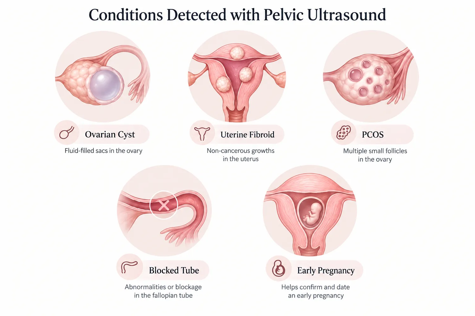
What Can a Pelvic Ultrasound Detect?
This scan helps identify a wide range of conditions, including:
- Ovarian cysts
- Uterine fibroids
- Endometrial thickness or abnormalities
- PMOS-related changes in the ovaries
- Blocked or fluid-filled fallopian tubes
- Early pregnancy and its location
- Structural issues affecting fertility
“A single scan can rule out or confirm so many possibilities. That’s why I don’t treat it as a routine formality. I look at it closely, every single time.” – Dr. Pooja Papishetty
Why Choose Matrika Fertility and Laparoscopic Centre for Your Pelvic Ultrasound?
| What We Offer | Why It Matters to You |
| In-house ultrasound facility | No running between labs; scan and consultation happen together |
| Scans reviewed personally by Dr. Pooja | The doctor treating you also reads your scan |
| Fellowship in Reproductive Medicine | Scans interpreted with a fertility specialist’s eye, not just a general reading |
| Same-day results and explanation | You leave with answers, not just a report |
| Located in Kondapur | Easy access for patients from Madhapur, Gachibowli, and Kothaguda |
How to Prepare for Your Pelvic Ultrasound
Preparation is simple and takes just a few minutes to get right.
- For a transabdominal scan, drink 3 to 4 glasses of water about an hour before your appointment and avoid urinating until after the scan
- For a transvaginal scan, an empty bladder is preferred, so no special water intake is needed
- Wear comfortable, loose clothing
- Carry any previous scan reports or prescriptions, if available
“Every scan tells a story. My job is to read it right and explain it to you in a way that actually makes sense.” – Dr. Pooja Papishetty, Matrika Fertility and Laparoscopic Centre